OBSTETRICS GYNECOLOGY AND IVF CLINIC
Doç.Dr.Yasemin Çekmez
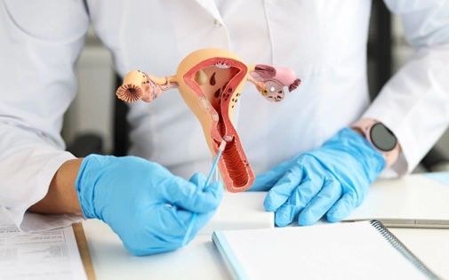
Our Services
Gynecologic Operations
Adres:
Kızkalesi sokak
Şua Elite Plaza
1CA
ŞERİFALİ - ÜMRANİYE
Türkiye
02163136662
Infertility
Gynecologic Diseases
Genital Aesthetics
Pregnancy follow up


ABOUT ME
I graduated from Malatya Scıence Hgh School ın 1999. Between 1999 and 2005, I graduated from Ankara Unıversıty Faculty of Medıcıne and receıved the tıtle of Doctor. Between 2006 and 2011, I receıved specıalızatıon traınıng ın Gynecology and Obstetrıcs at Goztepe Traınıng and Research Hospıtal. I RECEIVED IVF TRAINING AT ZEYNEP KAMİL HOSPITAL TRAINING AND RESEARCH HOSPITAL IN 2019. After workıng ın dıfferent traınıng and research hospıtals for many years as a Gynecologıst, I have been contınuıng to serve my patıents ın my prıvate clınıc ın umranıye sınce 2020.


OBSTETRICS GYNECOLOGY AND INFERTILITY CLINIC
Doç.Dr.Yasemin Çekmez
What Is Infertility (Sterility)
Infertility (sterility) is the condition, in which the couple have had sexual intercourse without contraception regularly for 1 year but they have not achieved pregnancy. If the woman is older than 35 years old, the waiting period to achieve pregnancy is reduced to 6 months. Infertility occurs in 15-20 out of every 100 couples and unfortunately occurs more frequently with increasing female age. In women older than 35 years old and in couples with a history of infertility (sterility), further investigations and appropriate treatment options should be started immediately
How Does Pregnancy Occur Naturally?

The ovaries are the main organs that contain female sexual (reproductive) cells. They contain millions of female reproductive cells that are too small to be seen with the naked eye or even with ultrasonography. These cells have a certain lifespan and gain different sizes and properties at each stage of their lives. Female eggs in the ovaries are called "follicles".
In the ovaries (right and left) of a healthy woman, in the first three days of menstruation, approximately 8-10 eggs (oocytes) develop into antral follicles so that they can be fertilized. Antral follicles are 2-7 mm in size and their size can be measured by ultrasound imaging. In order for these eggs to mature, the brain begins to secrete a hormone called FSH, and under the influence of this hormone, antral follicles begin to grow. One of these follicles grows faster and prevents the secretion of this hormone from the brain, preventing the other antral follicles from maturing further.

But; The first one, which contains the egg (oocyte), which is your dominant female sex cell selected for ovulation, will continue to mature. On the one hand, your selected egg secretes hormones that will prepare the inner lining of the uterus, increasing its thickness and making it suitable for the growth of the embryo. On the other hand, it continues to grow in a predetermined manner, increasing its size by 1-1.5 mm every day. Finally, this selected follicle cracks (ovulation) between the 13th and 15th days of the menstrual period and releases the female reproductive cell inside. This is actually a kind of little explosion that pushes the germ cell into the uterine tube. The hairy structures at the end of the tube catch the egg and take it inside.
The egg inside begins to move forward with the movements of the hair-like soft brooms inside the tube. Unfortunately, the lifespan of this egg is only 1 day. When the couple has sexual intercourse, male reproductive cells (sperm) are deposited at the entrance of the uterus (cervix). They will then begin to swim forward on their path using their own energy. First, they will reach the cervical canal, second, they will reach the uterine cavity, and then, moving through the tube, they will fertilize (fertilize) the female reproductive cell (egg or oocyte); He will only survive one day.

What cause infertility?
The causes of infertility may either belong to the man or the woman. However, no problems can be identified in 15% of the couples presenting for an evaluation. This is a condition called unexplained infertility, in which assisted reproductive techniques help achieve pregnancy. Also, there may be only one factor causing infertility or there may be more than one. The causes of infertility can be listed under two categories: male and female factors of infertility. The causes of infertility are distributed equally between the two genders at a rate of 40% in men and 40% in women in general. In the remaining 15-20% of couples, pregnancy cannot be achieved although no causes can be detected.
Women are born with an ovarian reserve of about 2 million eggs (oocytes), of which they will lose some of them each month until the end of their lifespans. There will be approximately 450,000 eggs left when they have their first menstrual bleeding. Approximately 1000 eggs will be dead each month before the woman is 35 years old, and about 1500 eggs will die every month after the woman becomes older than 35 years of age. How about doing a simple calculation? If we start by assuming that 1000 out of 450,000 eggs die each month, we can argue that you are likely to have 450 menstrual periods and you can get pregnant 450 times until menopause. In other words, a girl who begins to have her periods at the age of 12, will have menopause after 450 months (37 years) or at the age of 49 years, which is a reasonable age for menopause. With advancing age, the quality and fertilizability of the eggs decline, along with the reduction of their likelihood to adhere to the inner membrane of the uterus. In this respect, women over the age of 35 years and who have not got pregnant for 6 months should undergo a gynecologic evaluation as soon as possible. In men, the effect of age on fertility is not as distinct as in women.
Irregular menstrual cycles
Diminished ovarian reserve (low ovarian reserve)
Blocked fallopian tubes or adhesions inside the tubes
History of previous infections or intra-abdominal interventions
Endometriosis: It is described as the migration of the endometrium, which is the inner layer of the uterus and which is poured out with each menstrual bleeding every month, to another location in the body, impairing the functioning of the ovaries, uterus or the fallopian tubes.
High prolactin levels
Early menopause
Fibroids (myomas): Fibroids are benign tumors in the uterus. They hamper conception based on their location and size. When they are located especially inside the uterus, they can hinder implantation. When they locate out of the uterus, they can divert the tubes or they can cause blockage in the tubes by compressing them.
Intra-abdominal Adhesions: This term describes tissue adhesions inside the abdominal cavity following pelvic infections, appendicitis or abdominal or pelvic surgery.
Thyroid Problems: Too much or too little synthesis of the thyroid hormones by the thyroid gland may disrupt ovulation and cause infertility.
Cancer history and cancer treatment: In particular, cancers in the female reproductive system can lead to infertility. Radiotherapy and chemotherapy also have an act on the reproductive ability of women.
Other medical conditions: Disorders such as late puberty, amenorrhea (no menstrual periods), liver diseases or diabetes can cause female infertility.
Excessive caffeine consumption.
Being overweight
Smoking and alcohol use
Several diagnostic tests are carried out in couples presenting for the evaluation of infertility . The tests may reveal no underlying factors as it occurs in unexplained infertility or they may reveal more than one underlying factor.
What is early reduction of ovarian reserve? (premature ovarian failure)
Early depletion of the ovarian reserve It defines diminished ovarian functions before the age of 40. Anti-Müllerien hormone (AMH) is synthesized in the existing eggs in the ovaries and the levels of this hormone are used for making the diagnosis. AMH levels of <1><!--1-->
Especially patients with a history of ovarian surgery have a great risk of early reductionrgery due to a chocolate cyst (endometrioma), torsion, hemorrhagic cysts or cyst ruptures etc., and if they have not conceived for 6 months although they are under 35 years of age.
Early ovarian failure (premature ovarian failurof ovarian reserve. Individuals need to be consulted with an IVF specialist for reserve control if they have history of undergoing ovarian sue) may result from several genetic diseases. Especially patients with a family history of early menopause should have their ovarian reserves followed up regularly and they are advised to plan conception accordingly.
Early depletion of ovarian reserve may also develop in smokers, in individuals receiving cancer treatment (radiotherapy-chemotherapy), and in several medical conditions including obesity, vitamin D deficiency, insufficient intake of some antioxidants, or after exposure to various toxins.
After the diagnosis, treatment for infertility (IVF) should start immediately.
Early ovarian failure (premature ovarian failure) does not DEFINITELY mean EARLY MENOPAUSE . Just because you have regular periods, it does not mean that your ovarian reserve is good. Patients often think that their reserves are sufficient because they have their periods regularly. However, periods are often not irregular in poor ovarian reserve. Depletion of the ovarian reserve enough to disrupt the menstrual cycles indicates a considerable reduction in the likelihood of getting pregnant with IVF. Therefore, it will be prudent if women having the abovementioned risks immediately contact with an IVF specialist to have their ovarian reserve checked.
How can poor ovarian reserve be detected?
Every healthy female infant is born having an average of one and a half million eggs. At the time of menarche , adolescent girls have 400,000 to 500,000 eggs. Of these eggs, 1000 are lost every month, and in fact, a woman ovulates 400 - 500 times on the average during the reproductive period. Regarding the ovarian reserve and reproductive age, it is known that diminished ovarian reserve resulting in menopause occurs in one out of every 100 women before the age of 40 and in one out of every 10 women around the age of 45. Therefore, diagnostic tests to check and determine the ovarian reserve become primarily important.
The most effective method to measure the ovarian reserve is to test the Anti-Müllerian Hormone (AMH) levels and the second one on the rank is to count the number of follicles in the ovary (antral follicle count).
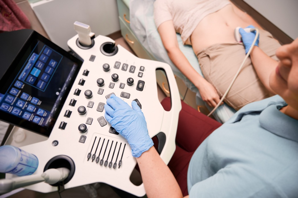
Transvaginal ultrasonography
The antral follicles count measured with ultrasound on the 3rd day of the menstrual period provides very reliable information to estimate the egg reserves. Presence of at least 6-7 potentially growing follicles (antral follicles) in the ovaries indicate a good ovarian reserve. However; if the number of follicles is less than 6-5, this will indicate a diminishing ovarian reserve. However, this finding should be evaluated in combination with other diagnostic tests, rather than to be used as a stand-alone method.
Measurement of the FSH (Follicle Stimulant Hormone) levels
Blood levels of FSH is tested on the 3rd day of the woman's menstruation. The test results provide information on the ovarian reserve. Synthesized by the pituitary gland in the brain, FSH stimulates the maturation of eggs in the ovaries. The main task of FSH is to stimulate the growth of the eggs and to induce estrogen hormone release. The better the woman's ovaries respond to this hormone, the less FSH will be secreted. After a decline in the number of eggs, consequently leading to lower levels of estrogen, FSH levels will be elevated again. If the FSH test on the 3rd day of menstruation reveals low levels, these levels will indicate good ovarian reserve. Higher levels will indicate diminished ovarian reserve. In this context, patients with FSH values of over 10 are at risk for having dinminished ovarian reserve.
AMH (Anti Müllerian Hormone)
When ovarian reserve is reduced, AMH levels will decline. The AMH test is a very reliable method for determining ovarian reserve. This test can be performed any day. Although the hormones FSH and LH act on the ovarian functions, they are the indirect markers to determine ovarian reserve. Because AMH is produced directly in the ovaries, it clearly indicates the ovarian reserve and ovarian functions.
Causes of Infertility in Men:
Unlike women, the causes of infertility in men are not many. In general, they include abnormalities in the count, shape, and the motility of the sperms.
Reduction in sperm count and motility
Structural abnormalities in sperms
No sperm cells in the semen (azoospermia)
Ejaculatory duct obstruction
Retrograde ejaculation (ejaculating into the bladder)
Hormonal causes
Undescended testicle at birth or after the birth
Feverish illnesses in childhood, diseases such as mumps in adolescence
Genetic diseases
Varicocele (dilation of vessels supplying and draining testis)
Giseases such as syphilis and gonorrhea
Sexual dysfunction (such as erectile disorders and premature ejaculation)
Diabetes
Previous cancer treatment
Infections
Testosterone deficiency
Smoking or alcohol use
Stress
Female external genital organs; It consists of the bony part under the navel, which we call mons pubis, the labia major (outer lips), the labia minor (inner lips), the clitoris (the upper junction of the labia minora), and the hymen, which we call hymen. Problems related to the sexual area can be examined in three ways: congenital, developmental and post-pregnancy problems. In some young women, the external anatomical structure may be underdeveloped, underdeveloped or asymmetrical. As the person begins to get to know his own body, differences in the genital area will cause discomfort.
Thanks to the advances in the field of aesthetic surgery, surgical and non-surgical aesthetic interventions in the female genital area have resulted in great developments. The aim here is to make the person feel good socially and to increase their harmony with their partner in their sexual life.
What does genital aesthetics include?
Genital aesthetics includes many procedures such as removing existing excess fat, increasing volume by filling or injection of your own fat, eliminating sagging, lightening the color, reducing the size of the inner lips, and narrowing the vagina. Genital aesthetics improves the genital muscles, narrows and reshapes the diameter of the vagina, returning it to its former narrow state. While vulvoplasty, labiaplasty or perineoplasty is an aesthetic procedure for the external appearance of the vagina, Vagionoplasty is for the internal structure of the vagina. Women generally have genital aesthetics performed due to their frequent complaints of sagging, which is related to the vaginal width and the size of the inner lips. By improving the aesthetic appearance of the genital area, the woman's self-confidence is increased and she is supported in enjoying sexual intercourse.
What are Genital Aesthetic Surgeries?
As we get older, the outer lips begin to tighten due to aging, that is, the fatty tissue begins to decrease. Apart from tightening the pubic area and the outer lips, removing fat from the upper part or performing fat injection, pubic aesthetics, reduction of the inner lips, labiaplasty, and vaginal tightening operations, which we call vaginoplasty, are some of the genital aesthetics that women need. In cases where thinning of the outer lips is observed, the necessary amount of fat tissue from the person's regional fat can be removed with the Liposuction technique and transferred to the thinned areas. A fuller and younger appearance can be achieved in the genital areas that appear saggy with fat transfer (fat graft application - fat injection) or filler applications.
what is Labiaplasty ?
The inner lips called "labium minor", which are generally congenital or grow, sag and become irregular with puberty, look bad aesthetically and take on an uncomfortable form, especially when sitting. Sometimes, one of the inner lips, which we call "labial asymmetry", may be longer or have a different structure than the other. Surgical correction of the inner lips is called Labioplasty. Deformations in the Labium Major (Outer Lip) or Labium Minor (Inner Lip) in the female external genital area can become sexually and socially disturbing. It is possible to correct these deformations with appropriate surgical approaches. Aesthetic surgery of the outer lips, called labia majora, is called vulvoplasty. After these interventions, which can be performed under local anesthesia or sedation, a 4-week sexual abstinence is applied. At the end of 4 weeks, the patient can have a healthier and more vibrant sexual life without any visible scars from the surgery.
what is Vaginoplasty ?
The vagina is one of our organs that is most prone to enlargement as a result of reasons such as advancing age, normal birth, sexual intercourse, abortion and similar interventions. Vaginal tightening surgery, also known as "Vaginoplasty", is performed to restore the enlarged vagina to its previous tight state. For this purpose, surgical tightening can be done, as well as laser tightening. With this new technique, application is performed without using a scalpel and faster recovery is achieved. The vagina may become deformed due to hereditary or loosening after birth. This problem, which negatively affects sexual life, is narrowed by removing the mucosa and muscle tissue from the back wall of the vagina and stitching it back together to narrow the vaginal canal. The patient can return to his/her daily life after a few days, and sexual abstinence is applied for approximately 4 weeks after the procedure. cliteroplasty The clitoris is the bulge located in the upper junction of both labia, just above the urinary canal. Sometimes being too big can cause discomfort and lack of self-confidence in women. Excess tissue is surgically removed, and the remaining part is sutured using dissolvable stitches. Cliteroplasty surgery can sometimes be combined with labiaplasty aesthetics.
Application of Liposuction as Vulva aesthetics
Starting from the lower part of the abdomen and continuing to the groin, the large area containing the "Pubis", the outer and inner lips, that is, the "major and minor labium" and the vagina, is the genital area. The biggest problem we encounter with the pubis is the sagging that occurs with the increase in fat tissue here and the deterioration of skin quality. This is an uncomfortable condition that appears as a swelling in the lower abdomen, especially when wearing trousers or a swimsuit. The solution is liposuction and stretching and recovery surgeries to reduce the fat tissue here. The bulge in the upper part of the female genital area (mons pubis) generally causes both sexual and social distress in patients with high body mass index. Socially, unwanted bulge/sagging in clothes is the most common cause of complaint. It is very easy to get rid of the swollen appearance thanks to liposuction applied on the vulva. Vulvoplasty genital aesthetics In the outer part of the genital area in women; Thinning of the tissues and sagging of the skin may be observed due to age and weight loss. We can regain the lost volume thanks to fat transfer taken from the patient or hyaluronic acid-based dermal fillers.
Non-surgical Applications in Female Genital Region
Dissolvable, ready-made filling materials are applied to the female genital area, non-surgically, in order to eliminate thinning and sagging, especially as a result of volume loss. The most commonly applied filling material is Hyaluronic Acid. This application can be performed in a clinical setting after the application of topical anesthetic cream. Lasers used in vaginal tightening are achieved by the recovery and renewal of collagen tissue by giving radiofrequency or ultrasound energies to narrow the vagina. These laser treatments, called non-surgical vaginoplasty, can be applied in a single session or in 3 sessions. Genital aesthetics Prices Genital aesthetic prices vary depending on the procedure performed. It is not legal for Ministry of Health-approved centers to list prices on their websites. You can get information about genital aesthetic prices by calling our polyclinic at +902122414624. To whom is vaginoplasty performed? It is especially performed on people with advanced age and postnatal deformities. It is also performed on people with urinary incontinence problems. We can briefly call the vaginoplasty procedure a tightening procedure. Who can undergo perinoplasty? During birth, an incision is made between the vagina and the anus area. After birth, sexual pleasure decreases because this incision does not heal completely or becomes large. Perinoplasty can also be performed together with vaginal tightening.
To whom is Labioplasty performed?
Some women may have deformities in the inner and outer lips of the vagina due to genetic characteristics. Labioplasty is performed to solve problems such as large vaginal outer lips, thickness and length disorders. Who Should Have Hoodoplasty? It is performed to correct disorders in skin folds. Hoodoplasty is performed especially on people with defects in the skin folds of the clitoris area or on people with a large clitoris.
What is Hymen Stitching?
It is the process of stitching the hymen that is torn due to situations such as sexual intercourse, impact or accident. This procedure is usually performed by people who have problems with virginity.
How is Genital Aesthetics Procedure Done?
A physical examination is performed before this procedure is performed. During physical examination, disorders in the genital area are determined. It is decided which method will be used to perform genital aesthetic surgery. Afterwards, the person's health history is listened to. After the necessary tests and controls are performed, if there is no obstacle to the procedure, genital aesthetics is performed. Local or general anesthesia is performed for genital aesthetics. Afterwards, the excess tissues determined during the physical examination in the vagina are removed. After the channel reduction process, stitching is done. Thus, the process ends. The type of operation and its progress may vary depending on which method will be applied. The operation time varies depending on which technique will be used.
After the procedure is completed, the person is kept under observation for the first 24 hours. If there are no problems at the end of the period, the patient is discharged.
What is the Recovery Period After Genital Aesthetics?
The recovery period may vary depending on which method of genital aesthetics was performed. It is normal for bruising, swelling or edema to occur after the operations. These complications are temporary. Using the medications prescribed by your doctor during the recovery period will minimize the feeling of pain. You should stay away from sexual intercourse and be careful not to use tampons or menstrual cups until the recovery period is over. You should be very careful about cleaning the genital area. You should choose your laundry from cotton laundry. Activities such as sports, heavy exercises and swimming should be postponed until after the recovery period.
Will There Be Any Scars After Genital Aesthetics?
Since the blood circulation in the genital area is rapid, the healing of the wound is better and faster. Therefore, there is no trace left. What Should You Pay Attention to After Genital Aesthetics? Genital hygiene is very important after aesthetics. You should choose looser clothes. The stitches will dissolve and dissolve on their own. After the operation, you should definitely use the antibiotics recommended by your doctor against genital area infections.
Will the dreamed aesthetic appearance appear after genital aesthetics?
During the physical examination performed before the operation, it is explained what the patient's appearance will be like when the existing problems are corrected. Most people are generally satisfied with the appearance that emerges after genital aesthetics.
How many days does it take to return to daily life after the operation? You can easily return to your daily life starting from the first 3-4 days.
Can genital aesthetics be performed during pregnancy? No, it cannot be done. It is quite dangerous. Can genital aesthetics be performed during breastfeeding? It is not appropriate to have any genital aesthetic surgery during breastfeeding and pregnancy.
What is the age limit for genital aesthetics? The age limit for aesthetic operations is +18. Is hymen stitching a genital aesthetic procedure? No it is not. It is generally performed in people whose hymen is completely closed, when it is necessary to open it for health reasons.
I
What is Genital Aesthetics?
Genital Aesthetic interventions are the name given to aesthetic methods used to eliminate acquired or congenital deformities in the genital area. While genital area deformities first and foremost create a lack of self-confidence, on the other hand, they negatively affect a woman's sexual life and daily life, and over time, can cause serious problems such as sexual reluctance, discharge or anxiety during intercourse.
The purpose of pregnancy follow-up is to protect the health of the mother throughout pregnancy and to ensure that the baby is born healthy. Pregnancy follow-up begins as soon as pregnancy is diagnosed. Approximately 2 weeks after pregnancy is diagnosed with blood and urine tests, the gestational sac can be seen with ultrasound. During this period, the status of ectopic pregnancy is checked by checking whether the gestational sac is inside the uterus. Approximately 1-2 weeks later, the baby's heartbeat can be detected with vaginal ultrasound. Blood and urine tests are then performed to determine the mother's general condition. It is determined whether there is a risk for diabetes, thyroid disease and blood incompatibility.
What should be the frequency of pregnancy follow-ups?
If the baby is single, follow-up every 2-3 weeks is appropriate for the first 3 months. Monthly follow-up is then continued. In cases of multiple pregnancies, existing illnesses in the mother, and recurrent miscarriages, follow-up is continued at frequent intervals. Here, the determining factors in the follow-up period are the risks and health status of the pregnant woman. If there are risk factors in the mother, such as the mother being older or younger, being too fat or too thin, having hypertension, diabetes, anemia and uterine abnormalities, follow-ups are carried out differently and more frequently.
Weekly checks during pregnancy
10-14. In the following weeks, the baby's neck thickness is checked with ultrasound. A double screening test is performed. Thus, early diagnosis of diseases such as Down Syndrome can be made.
16-18. A triple screening test is requested from the pregnant woman in the following weeks. The baby is evaluated with ultrasound. If the triple screening test is high, amniocentesis is performed to confirm the diagnosis. Amniocentesis is taking a sample of the baby's fluid with a needle. In experienced hands, the risk of amniocentesis is low.
20-24. Since the baby's organ development is largely completed in the following weeks, detailed ultrasonography is requested. In this ultrasound performed by the radiologist, attention is paid to all abnormalities in the baby.
24-28. A sugar challenge test is performed to detect gestational diabetes in the following weeks. Gestational diabetes is called "gestational diabetes". It poses a risk for the baby. Pregnant women whose sugar loading test is faulty are given an appropriate diet. Pregnant women with blood incompatibility are given blood incompatibility injections at the 28th week.
28-36. The baby is continued to be monitored with ultrasound between weeks. The mother is checked for urinary tract infection. The mother's weight and blood pressure are monitored.
After the 36th week, the gynecologist determines the mode of delivery. If there is an obstacle to normal birth, a cesarean section is planned. NST test is performed to evaluate the baby's condition. NST tests, which are performed once a week starting from the 36th week, are performed every 2-3 days after the 40th week.
In the last weeks, the amount of baby's water (amniotic fluid) is measured by ultrasound. After the 40th week, pregnancy is seen every 2-3 days. If labor does not begin, the pregnant woman is hospitalized and medications are given to initiate labor.
WHAT IS PREGNANCY MONITORING?
WHAT SHOULD WE CONSIDER FOR A HEALTHY PREGNANCY AND AFTER?
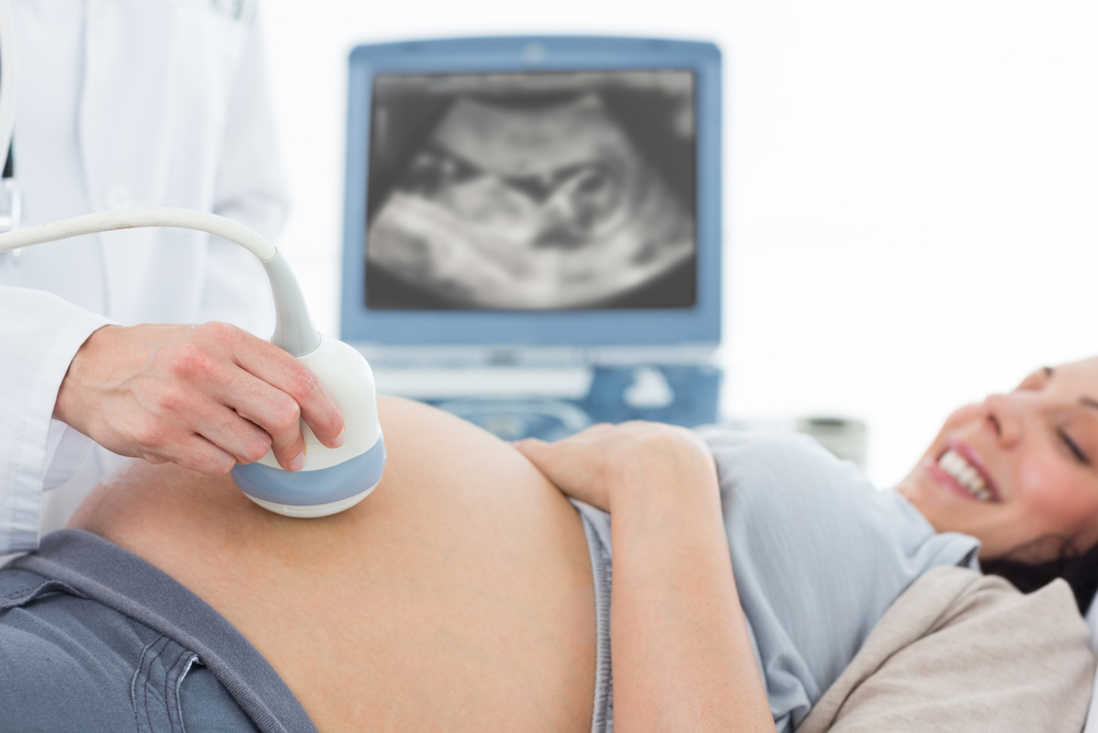
The purpose of pregnancy follow-up is to protect the health of the mother throughout pregnancy and to ensure that the baby is born healthy. Pregnancy follow-up begins as soon as pregnancy is diagnosed. Approximately 2 weeks after pregnancy is diagnosed with blood and urine tests, the gestational sac can be seen with ultrasound. During this period, the status of ectopic pregnancy is checked by checking whether the gestational sac is inside the uterus. Approximately 1-2 weeks later, the baby's heartbeat can be detected with vaginal ultrasound. Blood and urine tests are then performed to determine the mother's general condition. It is determined whether there is a risk for diabetes, thyroid disease and blood incompatibility.
![]()
Powered by estimated due date app
GYNECOLOGIC DISEASES
Gynecological diseases, which are medically called gynecological, are the main aim of the gynecologist doctor to get to the root of the disease that causes the complaint and to prolong the patient's quality of life and life by preventing the complaint.
Gynecology (Gynecology)
The main goal of the gynecologist is to prolong the patient's quality of life and life by getting to the root of the disease that causes gynecological complaints, medically called gynecological diseases, and preventing the disease. How long the disease has existed, how much it affects the patient, what increases or decreases the pain, multiple problems are investigated and the treatments applied are questioned. In obstetrics, questions such as how to have a healthy pregnancy, what to do, how to prepare for pregnancy, what are the risks that may be encountered are asked. or is important for patients preparing for pregnancy and needs to be answered.
What are Gynecological Diseases?
The most common gynecological diseases in women are:
Uterine diseases (myomas, adenomyosis, etc.).
Menstrual problems (menstrual irregularity, excessive menstruation, etc.).
Polycystic ovarian disease (PCOS),
Vaginal discharge, myomas, endometriosis,
Pelvic inflammatory disease or groin infection,
vaginitis,
Menopause,
Pain during sexual intercourse.
Uterine Diseases
The female uterus is the reproductive organ in which the pregnancy develops, is protected, and plays a role in pushing the fetus out with its contractions when the time comes. In its lower part, it is connected to the vagina with a structure called the cervix or cervix. The upper part forms the fundus. From this section, it opens to the ovaries with the help of bilateral fallopian tubes. It is attached to the hip bone by very strong ligaments from the cervix and is prevented from protruding. 8 out of 10 women face forwards and 2 out of 10 women face backwards. The condition in which the uterus is turned backwards, popularly known as inverted uterus, does not have any negative consequences for the patient. When the diseases of the uterus are examined, many problems that arise from birth of the uterus, the inner wall of the uterus, the outer wall of the uterus, or the loosening of the ligaments holding the uterus due to infection, come to the fore. To list these briefly:
myomas
adenomyosis
endometritis
Intrauterine
Polyps
hematometra
Congenital Uterine Abnormalities
Endometrial Hyperplasia
Endometrial Cancer
Uterine Sarcoma
Cervical Abnormalities
Cervical Cancer
Ovarian Diseases
myomas
Myomas are smooth muscle tumors of the uterus. These are the elements that most often cause problems in the uterus. It manifests itself with complaints such as bleeding, pressure and pain. Its treatment is laparoscopic, hysteroscopic myoma removal surgery. If it cannot be performed, open myoma surgery should be performed.
adenomyosis
It forms a myoma-like mass on the uterine wall. Its difference from myoma is that it is a tumor originating from endometriosis tissue. endometritis It is an infection of the inner wall of the uterus. Spiral is manifested by odorous discharge and fever after abortion or birth.
Intrauterine Polyps
It is recommended to remove tissue pieces growing inside the uterus hysteroscopically due to the risk of vaginal bleeding and harboring malignant cells. hematometra It is a condition in which menstrual blood accumulates inside the uterus as a result of not being able to be expelled. It occurs with intense groin pain.
Congenital Uterine Abnormalities
These abnormalities can be divided into many different groups and named differently. Some of these are double uterus, webbed uterus, and deformities called arcuate uterus. Normal anatomy is achieved through hysteroscopic and laparoscopic surgical interventions.
Endometrial Hyperplasia
It is a precursor to uterine cancer. It causes irregular vaginal bleeding. Especially the types carrying atypia carry a high risk of transformation into cancer. Depending on the patient's age and type of hyperplasia, biopsy follow-up, hormonal intrauterine device or uterine removal options may be applied.
Endometrial Cancer
These are tumors originating from the part of the uterus that creates menstrual bleeding, also called uterine cancer. Early diagnosis and surgical treatment are important. Depending on the stage, surgery is performed to remove the expanded uterus, ovaries and surrounding lymph nodes.
Uterine Sarcoma
It is a tumor originating from the wall of the uterus. Surgery and, if necessary, additional radiotherapy are applied.
Cervical Abnormalities
Evaluation of the cervix with smear helps in the early diagnosis of cervical abnormalities that may turn into cervical cancer. Since HPV is the most important cause of cervical cancer, it is important to perform HPV typing during the examination. Depending on the patient's smear and HPV results, colposcopy and biopsy are recommended if necessary.
Cervical Cancer
Cervical cancer is a preventable cancer. A woman who has regular check-ups can be diagnosed early and taken under control with a conization operation. Trachelectomy, which is a reproductive and uterus-preserving cervix removal surgery, can be performed in young patients. In advanced cases, radical cervical cancer surgery is performed. In cervical cancer, which has many pathological stages, surgery or radiotherapy is decided according to the staging. Pathological stages are not given on this site for patient information purposes.
Ovarian Diseases
If we examine the common ovarian diseases, ovarian cysts and tumors come to the fore. While simple cystic diseases of the ovary and polycystic ovaries are common, it is important to diagnose congenital ovarian tumors such as dermoid cysts, complicated diseases such as chocolate cysts and malignant tumors of the ovary early. Ovarian cysts often present as silent cystic masses. It is detected when it grows excessively, causes abdominal swelling, causes pain, and is detected during ultrasound examination. Complaints of urgent abdominal pain may require consulting a doctor. The rupture of the cyst and its contents spilling into the intra-abdominal membrane may cause severe abdominal pain. This cyst may have a simple structure, a chocolate cyst (endometrioma) or a dermoid cyst. Bleeding into the cyst often manifests itself with intense abdominal pain. Rarely, bleeding is so intense that it can lead to a drop in blood pressure and a life-threatening condition called hypovolemic shock. Torsion is the rotation of the ovarian stalk and disruption of its nutrition, depending on the size of the cyst. It is an emergency. If left untreated, it will result in loss of the ovary. Ovarian tumors can be benign or malignant. While benign forms are observed as serous or mucinous cystadenoma, very slowly developing intermediate forms are called borderline. Malignant forms can be called serous cystadenocarcinoma, mucinous cystadenocarcinoma, endometrioid cancer, clear cell cancer, sex cord stromal tumor, teratoma, dysgerminoma. Early symptoms may include loss of appetite, fatigue, fluid accumulation in the abdomen, a mass in the groin, and high blood Ca125 levels.
Menstrual Problems
The most common problems experienced by women regarding menstruation are as follows:
Irregular period
Excessive Menstrual
Bleeding Cessation of Menstruation (Amenorrhoea)
Painful Menstruation
Irregular period Heavy menstrual bleeding, intermenstrual bleeding, bleeding more than 23 days later than 35 days, bleeding lasting more than 7 days, bleeding after intercourse, amenorrhea, post-menopausal menstruation and painful menstruation are considered menstrual irregularities. Excessive Menstrual Bleeding In most women who experience heavy menstrual bleeding, no pathological findings are detected. These are cases where bleeding cannot be stopped due to hemostasis abnormalities of the endometrium in the uterine tissue or intrauterine prostaglandin abnormalities. It can be controlled with various medications. The main cause of bleeding in 30% of women is myoma. Myoma is a benign smooth muscle tumor located in the uterus. Endometrial Polyps, which are caused by intrauterine tissue growth, occur in 10% of women. Chronic pelvic infection, ovarian tumors, endometrial and cervical cancers can often cause vaginal bleeding. Thyroid diseases, hemostatic diseases such as von Willebrand and anticoagulant therapy may cause increased menstrual bleeding.
Menstrual Cessation
Menstrual cessation is divided into 2 groups. While the absence of menstruation until the age of 16 is called primary amenorrhea, the situation where the menstruating woman stops menstruating for more than 6 months is called secondary amenorrhea. The most common condition in which primary amenorrhea occurs is delayed puberty. There are also cases of inability to menstruate due to cervical stenosis, closed hymen or vaginal veil. The cause of secondary amenorrhea may be the possibility of pregnancy in the reproductive age, polycystic ovary syndrome, hyperprolactinemia or early menopause.
Painful Menstruation
Painful onset of menstrual periods in a woman entering reproductive age is called primary dysmenorrhea, and when normal painless menstruations turn into painful ones with advancing age, it is called secondary dysmenorrhea. Primary dysmenorrhea is associated with uterine contractions and decreased blood supply to the uterus due to increased prostaglandin levels in the endometrium. Secondary dysmenorrhea is often associated with groin area problems. The most common cause is myomas, adenomyosis, endometriosis or chocolate cyst disease, groin infection or ovarian cysts and tumors.